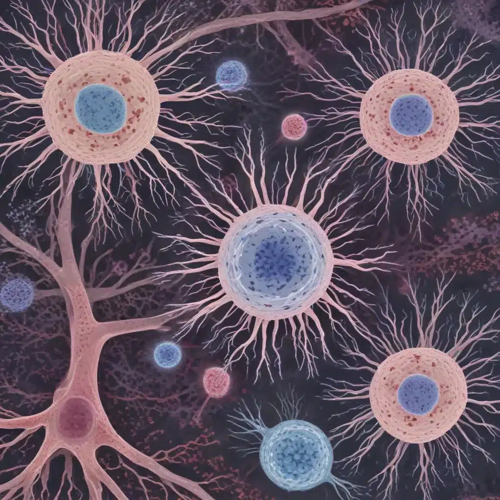
The endoplasmic reticulum (ER) is a central hub of diverse cellular processes, from protein synthesis to lipid metabolism. However, age-related dysfunction of ER-mediated pathways is a hallmark of many chronic diseases. Understanding the mechanisms underlying ER changes during aging is crucial for promoting healthier longevity. Recent research reveals that the morphological dynamics of the ER itself play a vital, yet underappreciated, role in modulating cellular and organismal aging.
Role of Autophagy in ER Remodeling
The ER is not a static organelle; rather, it continuously remodels its intricate network of sheet-like rough ER and tubular smooth ER subdomains to match the functional demands of the cell. This structural plasticity is driven by the relative abundance of ER-shaping proteins, such as reticulons and translocon subunits.
An alternative pathway for ER remodeling involves the selective turnover of ER components via a process called ER-phagy. ER-phagy can occur through both microautophagy (direct lysosomal engulfment) and macroautophagy (packaging into autophagosomes). Intriguingly, ER-phagy pathways often intersect with longevity-promoting interventions, suggesting this selective autophagy may play a critical role in aging biology.
Age-Dependent Changes in ER Structure
Using advanced microscopy techniques in the model organism Caenorhabditis elegans, researchers have uncovered profound, age-onset remodeling of ER networks. As adult worms age, the ER undergoes a dramatic shift from densely packed, rough ER sheets to a more diffuse, tubular network. This remodeling is accompanied by a substantial decline in total ER volume and mass, particularly of the rough ER subdomains.
Importantly, these age-dependent ER alterations occur across diverse cell types, including the intestine, muscle, and neurons. The universal nature of these ER changes suggests they may represent a fundamental cell biological hallmark of the aging process, potentially underpinning the widespread ER dysfunction observed in age-related diseases.
Mechanisms of ER-phagy
To drive this substantial ER remodeling, the ER-phagy program is activated early in adulthood. This selective autophagy pathway targets rough ER subdomains, likely in response to rises in luminal protein-folding burden and reductions in global protein synthesis rates – hallmarks of cellular aging.
Intriguingly, impairing ER-phagy limits lifespan in yeast, mirroring the effects of blocking general macroautophagy. Conversely, diverse longevity-promoting interventions, such as reduced insulin/IGF-1 signaling or mTOR inhibition, induce profound remodeling of ER morphology even in young animals. These findings indicate that ER remodeling via ER-phagy is an adaptive, protective response during aging.
ER Function and Homeostasis
The ER houses numerous essential cellular processes, from protein quality control to lipid metabolism and calcium signaling. The ER’s central positioning in cell and organismal physiology means that disruption of ER homeostasis can trigger widespread dysfunction, from the subcellular to the organismal scale.
Age-related declines in ER proteostasis, such as reduced chaperone levels and impaired stress signaling (the unfolded protein response), are well-documented. However, the morphological remodeling of the ER itself represents an underappreciated mechanism by which aging can shape ER functionality.
Cellular Stress and ER Stress Response
The ER’s different subdomains are optimized for distinct metabolic tasks. Rough ER sheets, enriched in ribosomes and protein folding machinery, are tailored for proteostasis. In contrast, the tubular smooth ER is more involved in lipid metabolism and organelle interactions.
As the ER shifts towards a more tubular morphology with age, its proteostasis capacity declines while lipid-related functions may be maintained or even enhanced. This remodeling likely contributes to the metabolic reprogramming often observed during aging, where cellular metabolism shifts away from protein synthesis and towards lipid metabolism.
ER-Associated Degradation (ERAD)
The ER also plays a crucial role in protein quality control through the ER-associated degradation (ERAD) pathway. ERAD identifies and retrotranslocates misfolded proteins from the ER lumen to the cytosol for proteasomal degradation.
The age-dependent loss of rough ER sheets may compromise ERAD efficiency, leading to the accumulation of damaged proteins that can trigger chronic ER stress responses and broader cellular dysfunction.
Regulation of ER Dynamics
The structural and functional specialization of ER subdomains is dynamically regulated by an intricate network of ER-shaping proteins, chaperones, and interactions with other organelles. Understanding how this delicate ER homeostasis is perturbed during aging may reveal new therapeutic avenues for targeting age-related diseases.
Cellular Aging and ER Remodeling
Senescence and ER Morphology
As cells undergo cellular senescence, a state of stable cell cycle arrest often associated with aging, the ER also undergoes substantial remodeling. Senescent cells exhibit a shift towards a more tubular ER network, mirroring the age-dependent changes observed in healthy tissues.
This ER remodeling in senescent cells may contribute to the characteristic secretory phenotype, where senescent cells release a plethora of inflammatory cytokines, growth factors, and proteases that can disrupt tissue homeostasis.
Age-Onset ER Alterations
The profound, age-dependent remodeling of the ER appears to be an early and progressive event in the aging process, preceding many other well-characterized hallmarks of aging. This suggests the ER may act as an integrator and driver of diverse age-related cellular changes.
Implications for Tissue Homeostasis
The widespread nature of age-dependent ER remodeling, occurring across multiple cell types and tissues, implies that disruption of ER structure-function relationships is a unifying mechanism underlying tissue dysfunction during aging. Targeting the pathways governing ER dynamics may therefore represent a promising, holistic approach to promoting healthier longevity.
Impact of ER-phagy on Organismal Aging
Longevity and ER Remodeling
Interestingly, multiple longevity-extending interventions, such as reduced insulin/IGF-1 signaling, mTOR inhibition, and translation suppression, all induce profound remodeling of ER morphology even in young animals. This suggests that the ER-phagy-driven remodeling of ER networks may be a conserved, adaptive response that promotes healthy longevity.
Therapeutic Targeting of ER-phagy
Given the crucial roles of the ER in cellular homeostasis and the potential links between ER remodeling and longevity, pharmacological modulation of ER-phagy pathways represents an intriguing therapeutic strategy. Selectively enhancing ER-phagy to promote healthy ER structure-function relationships may help mitigate age-related diseases.
Evolutionary Perspectives on ER Dynamics
From an evolutionary perspective, the pronounced age-dependent remodeling of the ER may represent a double-edged sword. While the initial ER remodeling in early adulthood appears to be a protective response, the progressive and unrestrained loss of rough ER in later life may ultimately contribute to the various pathologies of aging.
This highlights the concept of antagonistic pleiotropy, where traits that are beneficial early in life can become detrimental later on. Understanding the nuanced roles of ER dynamics in aging may provide important insights into the fundamental evolutionary drivers of the aging process.
In conclusion, the endoplasmic reticulum is an increasingly appreciated hub of cellular aging biology. The morphological remodeling of ER networks, driven by ER-phagy, represents a previously underappreciated mechanism by which the ER shape and function change with age. Targeting these ER dynamics may yield new avenues for promoting healthier longevity. For the latest insights and design inspiration, be sure to visit https://www.reluctantrenovator.com.


